Trisomy 18, the second most common autosomal trisomy after Down syndrome, affects approximately 1 in 6,000–8,000 live births. When addressing this sensitive topic, it is important to understand that it is a rare genetic condition with significant consequences. Unfortunately, statistics show that about 50% of affected newborns do not survive beyond the first two weeks of life, and only one in five reaches three months.
Also known as Edwards syndrome, this condition is caused by an error in chromosome separation, most often of maternal origin. In fact, the risk increases with advancing maternal age, with higher incidence among women over 35. Additionally, trisomy 18 predominantly affects female fetuses, likely due to selective miscarriage. In this article, we will explore the signs, symptoms, and support options available for mothers facing this diagnosis during pregnancy.
What Is Trisomy 18 and Why It Matters During Pregnancy
Edwards syndrome is a severe chromosomal abnormality characterized by the presence of an extra chromosome 18 in the cells. This complex genetic condition requires special attention during pregnancy because of its profound impact on both the fetus and the parents.
Definition of Edwards Syndrome
Trisomy 18 is a genetic disorder caused by the presence of a third copy of chromosome 18. Normally, humans have 46 chromosomes (23 pairs), but individuals with this syndrome have 47. The condition was first described in 1960 by Dr. John Hilton Edwards.
Trisomy 18 is one of the few aneuploidies compatible with life, although it carries severe consequences. The chromosomal error usually occurs during the formation of reproductive cells (before conception) or during the first divisions of the fertilized egg. The extra chromosome almost always comes from the mother, and advanced maternal age significantly increases the risk.
Incidence and Impact on Pregnancy
Trisomy 18 is the second most common autosomal trisomy after Down syndrome, with an incidence of about 1 in 6,000–8,000 live births. When including miscarriages and pregnancy terminations due to fetal anomalies, the true frequency is estimated at about 4.1 per 10,000 pregnancies.
More than 95% of affected fetuses die in utero. Among live births, approximately 50% do not survive beyond two weeks, and only 5–10% reach one year of age. Survival into adulthood is extremely rare, although slight improvements have been observed in recent years.
A marked female predominance exists among live-born infants, with a female-to-male ratio of about 3:1, likely due to higher intrauterine mortality in male fetuses.
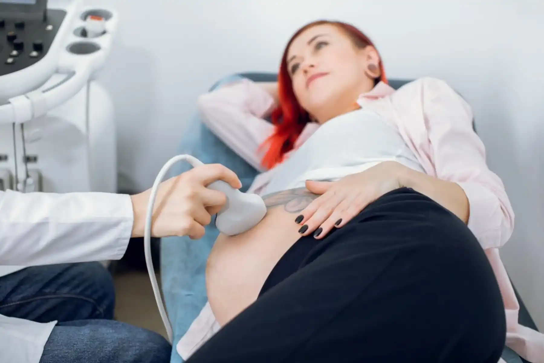
During pregnancy, trisomy 18 may be suspected based on ultrasound findings such as intrauterine growth restriction, multiple malformations, and choroid plexus cysts. Maternal serum markers may also be altered. Modern NIPT (Non-Invasive Prenatal Testing) offers a non-invasive screening option, although sensitivity and specificity are slightly lower than for trisomy 21.
Differences Between Complete, Mosaic, and Partial Trisomy 18
Trisomy 18 presents in three main forms:
- Complete trisomy 18: Accounts for about 94–95% of cases. All cells contain the extra chromosome and prognosis is generally poor.
- Mosaic trisomy 18: Occurs in about 5% of cases. Only some cells are affected, leading to variable severity.
- Partial trisomy 18: The rarest form (about 2%), caused by duplication of only part of chromosome 18, often due to a translocation.
Understanding these differences is essential for genetic counseling, as recurrence risk may be higher in families with chromosomal translocations. Overall recurrence risk for trisomies 13, 18, and 21 is about 1%.
Signs and Symptoms of Trisomy 18 During Pregnancy
Ultrasound Findings
Prenatal ultrasound is crucial for early suspicion of trisomy 18. The morphology scan around the 20th week can reveal:
- Intrauterine growth restriction (IUGR)
- Choroid plexus cysts
- Brain malformations (agenesis of the corpus callosum, brachycephaly)
- Clenched hands with overlapping fingers
- Omphalocele
- Single umbilical artery
Other findings may include kidney and intestinal anomalies.
Reduced Fetal Activity
Fetuses with trisomy 18 often show reduced movement, which may indicate fetal distress and prompt further investigation.
Polyhydramnios and Small Placenta
Polyhydramnios (excess amniotic fluid) is common and often associated with a small placenta. In some cases, oligohydramnios (reduced fluid) may occur, highlighting the importance of regular monitoring.
Cardiac and Brain Malformations
About 90% of affected fetuses have congenital heart defects, including ventricular and atrial septal defects, patent ductus arteriosus, tetralogy of Fallot, and aortic coarctation. Brain anomalies may include corpus callosum agenesis, choroid plexus cysts, microcephaly, or, in rare cases, anencephaly.
Prenatal Diagnosis: How Trisomy 18 Is Identified
Ultrasound Screening and Nuchal Translucency
Between 11 and 13+6 weeks, increased nuchal translucency is found in about 75% of affected fetuses. Absence of the nasal bone is also common.
Combined Test and Triple Test
- Combined test (first trimester): Combines nuchal translucency with maternal blood markers (PAPP-A and free β-hCG); sensitivity ~85%.
- Triple test (second trimester): Measures AFP, unconjugated estriol, and hCG; results are altered in trisomy 18.
These tests provide risk estimates, not definitive diagnoses.
NIPT: What It Is and How It Works
NIPT analyzes fetal DNA fragments in maternal blood from the 10th week onward. For trisomy 18, sensitivity is approximately 98%, with a very low false-positive rate. However, it remains a screening test.
Amniocentesis and Chorionic Villus Sampling
These invasive procedures provide definitive diagnosis. CVS is performed between weeks 10–12, and amniocentesis between weeks 15–18, with a miscarriage risk of 0.5–1%.
After Diagnosis: Options and Support
Genetic Confirmation and Counseling
Karyotype analysis confirms the diagnosis. Genetic counseling helps families understand recurrence risks, prognosis, and future reproductive options.
Therapeutic and Palliative Care
There is no curative treatment. Management focuses on supportive and palliative care, including feeding support, cardiac management, physiotherapy, and infection control. Pediatric palliative care plays a key role in maintaining quality of life.
Multidisciplinary Care Team
Care involves geneticists, neonatologists, cardiologists, neurologists, therapists, and pediatricians, aiming whenever possible to allow home care and family-centered support.
Psychological Support for Parents
Psychological support is essential. Associations such as SOFT Italia provide counseling, peer support, and guidance throughout diagnosis and beyond.
Living Pregnancy With a Trisomy 18 Diagnosis
Emotional Management and Family Communication
Shock, fear, and grief are common. Many parents report that, despite profound difficulty, the experience brings a deeper perspective on life. Studies show that 40–85% of couples offered pediatric palliative care choose to continue the pregnancy.
Support Associations
Organizations such as SOFT Italia and Fondazione Il Cuore in una Goccia support families by offering information, connection, and advocacy.
Rights and Protections in Italy
In Italy, trisomy 18 is classified as a rare disease (code RNG080) with full healthcare exemption. Law 104/92 provides paid leave, workplace protections, and disability benefits for families.
Conclusion
Facing a trisomy 18 diagnosis during pregnancy is one of the most challenging experiences a family can endure. Modern screening tools such as NIPT allow early, highly accurate detection from the 10th week of pregnancy. However, each family’s journey is unique.
What clearly emerges is the critical importance of multidisciplinary medical care, psychological support, and social assistance. Families are not alone, and support networks, healthcare professionals, and legal protections can help navigate this difficult path with dignity and informed choice.
Whatever decision parents make—continuing or terminating the pregnancy—it must be respected, supported, and guided by accurate information, compassion, and care.